What are PROTACs and How Do They Treat Diseases?
PROTACs and toxic protein removal
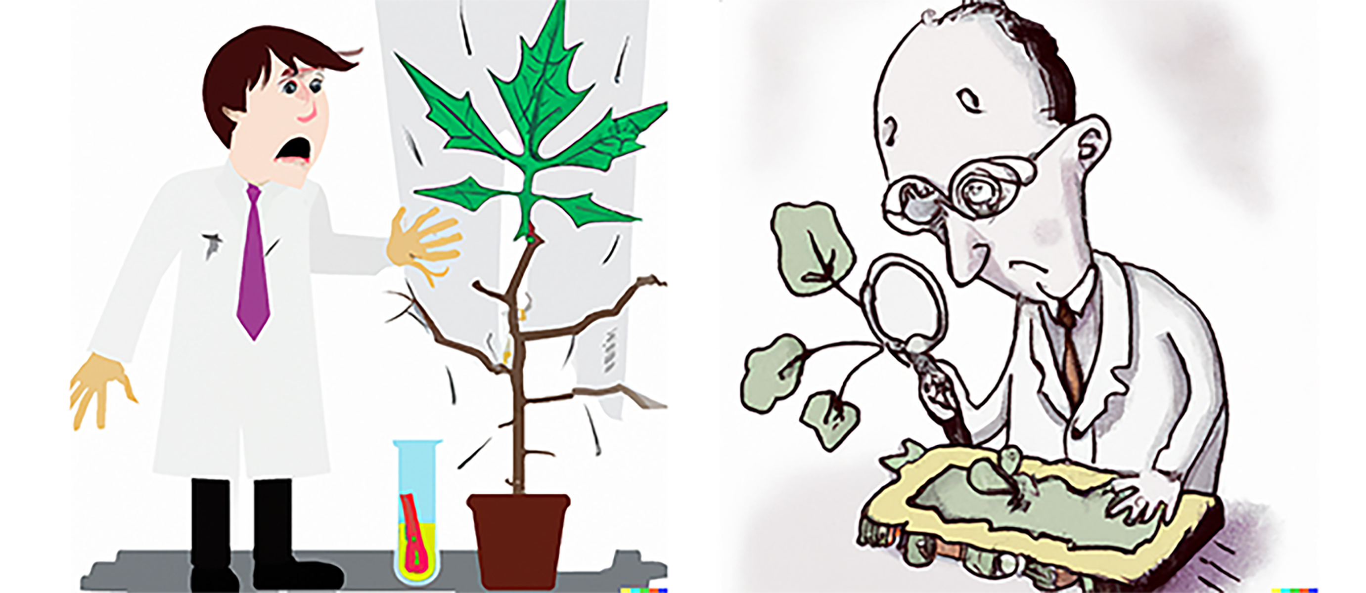
Protocol to extract plant proteins efficiently
Proteomics has become an effective tool for investigating plant functional genomics, allowing for the detection of post-transcriptional modifications. Two-dimensional gel electrophoresis (2-DE) has been a successful method for studying complex gene expression at the protein level. However, sample preparation for high-quality proteomic analysis can be difficult, especially when the proteins come from plant tissues that contain abundant metabolites and fewer proteins.

Given that the method of protein extraction used for plant samples can significantly affect the subsequent experimental results, we introduce here a rapid and universally applicable protocol for protein extraction from recalcitrant tissues, which is effective for the extraction of proteins from a variety of plant species. The protocol was created to quickly handle small amounts of tissue samples within an hour using microtubes. It can be increased in scale to handle more sizeable samples, such as 1 to 5 grams of tissue samples in 30 to 50 mL centrifugation tubes.
Protocol:

The use of a phenol/SDS mixture for protein extraction is more efficient than using either one of the buffers alone. This method has been employed successfully on a range of leaves and fruits and is a great choice for extracting proteins from delicate plants rapidly. This makes it a perfect choice for regular proteomic analysis.
PROTACs and toxic protein removal
Recent findings from Northwestern University using the Bioss PPAR Gamma Polyclonal Antibody and the Bioss YWHAZ (1B6) Monoclonal Antibody shed new...
A proposed role for the SARS‐CoV‐2 nucleocapsid protein in the formation and regulation of biomolecular condensates